135 results filtered with: Digital Images
- Digital Images
- Online
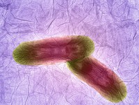
Nanographene oxide interacting with bacteria, TEM.
Suffian, Izzat.Date: 2015
- Digital Images
- Online
Astrocytes and blood vessels of the retina, micrograph.
Luna, Gabriel.Date: 2012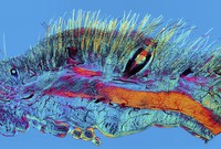
- Digital Images
- Online
Cat skin showing hairs, a whisker and their blood supply.
Linstead, David.Date: 2014
- Digital Images
- Online
Human stem cell embedded in a 3D matrix, Cryo SEM.
Ferreira, Silvia A.Date: 2015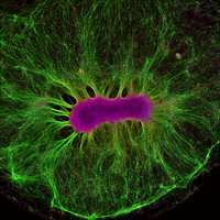
- Digital Images
- Online
Brain Organoid.
Edington, Collin.Date: 2017
- Digital Images
- Online
The Placenta Rainbow : immune system regulation of placental development, mouse.
Nadkarni, Suchita.Date: 2016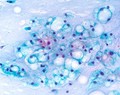
- Digital Images
- Online
Papanicolaou stained smear of a clival chordoma, microscopy. Chordomas are cancers formed of cells which resemble those of the notochord (spine) of a developing foetus. Although they can present anywhere within the spine and skull, the majority grow in the sacral region of the spine, corresponding to the lower back. This image shows a Papanicolaou (Pap) stained smear obtained from a needle biopsy of a chordoma in the clivus, a part of the cranium at the base of the skull.
William R. Geddie
- Digital Images
- Online
Adams' Universal Compound Microscope, late 18th century.
Adams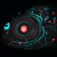
- Digital Images
- Online
Neuromast and neuron development, zebrafish.
Lekk, Ingrid.Date: 2015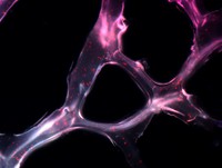
- Digital Images
- Online
Artificial microRNA scaffold.
Condé, João.Date: 2015
- Digital Images
- Online
Adams' Universal Compound Microscope, late 18th century.
Adams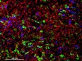
- Digital Images
- Online
Cellular architecture of human skin lymphoma imaged by whole mount tissue microscopy. Normal human skin has a rich network of white blood cells (specifically dendritic cells, T cells and macrophages) which form sheaths around blood vessels. In diseased skin, such as in skin lymphoma as seen here, this normal architecture becomes distorted. In this image, lots of T cells (stained for CD3; red), dendritic cells (stained for CD11c; green) and macrophages (stained for LYVE-1; blue) have infiltrated the skin. X20 magnification. Scale bar (white) represents 100 micrometres.
Dr. Xiao-nong Wang, Human Dendritic Cell Laboratory, Newcastle University
- Digital Images
- Online
Papanicolaou stained smear of a C2 vertebral chordomal mass, microscopy. Chordomas are cancers formed of cells which resemble those of the notochord (spine) of a developing foetus. Although they can present anywhere within the spine and skull, the majority grow in the sacral region of the spine, corresponding to the lower back. This image shows a Papanicolaou (pap) stained smear obtained from a needle biopsy of a chordoma of the C2 vertebrae, located at the top of the neck just underneath the base of the skull.
William R. Geddie
- Digital Images
- Online
Cross-section through a cluster of maize leaves, LM.
Federici, Fernán.Date: 2015
- Digital Images
- Online
Cellular architecture of normal human skin imaged by whole mount tissue microscopy. Human skin has a rich network of white blood cells (specifically dendritic cells, T cells and macrophages) which form sheaths around blood vessels. In this image, T cells (stained for CD3; red) dendritic cells (stained for MHC class II; green) and macrophages (stained for LYVE-1; blue with some cells showing a tinge of green) can be seen. Cell nuclei have been stained with DAPI (grey). This normal cellular architecture is grossly disrupted in diseased skin (see related images). X10 magnification. Scale bar (white) represents 200 micrometres.
Dr. Xiao-nong Wang, Human Dendritic Cell Laboratory, Newcastle University
- Digital Images
- Online
Cellular architecture of normal human skin imaged by whole mount tissue microscopy. Human skin has a rich network of white blood cells (specifically dendritic cells, T cells and macrophages) which form sheaths around blood vessels. In this image, T cells (stained for CD3; red) dendritic cells (stained for MHC class II; green) and macrophages (stained for LYVE-1; blue with some cells showing a tinge of green) can be seen. Cell nuclei have been stained with DAPI (grey). This normal cellular architecture is grossly disrupted in diseased skin (see related images). X20 magnification. Scale bar (white) represents 100 micrometres.
Dr. Xiao-nong Wang, Human Dendritic Cell Laboratory, Newcastle University
- Digital Images
- Online
Cellular architecture of normal human skin imaged by whole mount tissue microscopy. Human skin has a rich network of white blood cells (specifically dendritic cells, T cells and macrophages) which form sheaths around blood vessels. In this image, blood vessels (string-like structures stained for CD31; red), lymphatic vessels (ribbon-like structures stained for LYVE-1; blue) and dendritic cells (stained for CD11c; green) can be seen. Macrophages (stained for LYVE-1; blue) are also present. This normal cellular architecture is grossly disrupted in diseased skin (see related images). X10 magnification. Scale bar (white) represents 200 micrometres.
Dr. Xiao-nong Wang, Human Dendritic Cell Laboratory, Newcastle University
- Digital Images
- Online
Mouse embryonic posterior neuropore, confocal image.
Galea, Gabriel.Date: 2016
- Digital Images
- Online
Calvarial (skull) osteocytes, murine, THG and SHG
Daniela Malide, NIH, Bethesda, USA
- Digital Images
- Online
Cellular architecture of normal human skin imaged by whole mount tissue microscopy. Human skin has a rich network of white blood cells (specifically dendritic cells, T cells and macrophages) which form sheaths around blood vessels. In this image, blood vessels (string-like structures stained for CD31; green), lymphatic vessels (ribbon-like structures stained for LYVE-1; blue) and T cells (stained for CD3; red) can be seen. T cells are only found around dermal blood vessels. Macrophages (stained for LYVE-1; blue) are also present. This normal cellular architecture is grossly disrupted in diseased skin (see related images). X10 magnification. Scale bar (white) represents 200 micrometres.
Dr. Xiao-nong Wang, Human Dendritic Cell Laboratory, Newcastle University
- Digital Images
- Online
Cellular architecture of normal human skin imaged by whole mount tissue microscopy. Human skin has a rich network of white blood cells (specifically dendritic cells, T cells and macrophages) which form sheaths around blood vessels. This image was taken greater than 150 micrometres beneath the junction that joins the dermal and epidermal layers of the skin (dermo-epidermal junction). At this level, dendritic cells (stained for CD11c; green) and macrophages (stained for LYVE-1; blue) form clusters around blood vessels (stained for CD31; red). This normal cellular architecture is grossly disrupted in diseased skin (see related images). Scale bar (white) represents 100 micrometres.
Dr. Xiao-nong Wang, Human Dendritic Cell Laboratory, Newcastle University
- Digital Images
- Online
TEM - myelin sheath - detailed structure
Mike Kayser
- Digital Images
- Online
TEM - myelin sheath - detailed structure
Mike Kayser
- Digital Images
- Online
Normal bovine sperm
Paul Sharpe
- Digital Images
- Online
Normal bovine sperm
Paul Sharpe